
The human brain is a complex organ that allows us to think, move, feel, see, hear, taste, and smell. It controls our body, receives information, analyzes information, and stores information (our memories).
The brain produces electrical signals, which, together with chemical reactions, let the parts of the body communicate. Nerves send these signals throughout the body. SIZE OF THE HUMAN BRAIN
 The brain consists of gray matter (40%) and white matter (60%) contained within the skull. Brain cells include neurons and glial cells. The brain has three main parts: the cerebrum, the cerebellum, and the brain stem (medulla).
 NOURISHMENT OF THE BRAIN
Although the brain is only 2% of the body's weight, it uses 20% of the oxygen supply and gets 20% of the blood flow. Blood vessels (arteries, capillaries, and veins) supply the brain with oxygen and nourishment, and take away wastes. If brain cells do not get oxygen for 3 to 5 minutes, they begin to die. Cerebrospinal fluid (CSF) surrounds the brain. THE NERVOUS SYSTEM
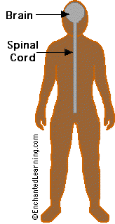
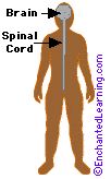 The brain and spinal cord make up the central nervous system (CNS). The brain is connected to the spinal cord, which runs from the neck to the hip area. The spinal cord carries nerve messages between the brain and the body. The nerves that connect the CNS to the rest of the body are called the peripheral nervous system. The autonomic nervous system controls our life support systems that we don't consciously control, like breathing, digesting food, blood circulation, etc.
 PROTECTION
The cells of the nervous system are quite fragile and need extensive protection from being crushed, being infected by disease organisms, and other harm. The brain and spinal cord are covered by a tough, translucent membrane, called the dura mater. Cerebrospinal fluid (CSF) is a clear, watery liquid that surrounds the brain and spinal cord, and is also found throughout the ventricle (brain cavities and tunnels). CSF cushions the brain and spinal cord from jolts.
 The cranium (the top of the skull) surrounds and protects the brain. The spinal cord is surrounded by vertebrae (hollow spinal bones). Also, some muscles serve to pad and support the spine.
 More subtly, the blood-brain barrier protects the brain from chemical intrusion from the rest of the body. Blood flowing into the brain is filtered so that many harmful chemicals cannot enter the brain.
The brain has three main parts, the cerebrum, the cerebellum, and the brain stem. The brain is divided into regions that control specific functions.
THE CEREBRUM:
Frontal Lobe
- Behavior
- Abstract thought processes
- Problem solving
- Attention
- Creative thought
- Some emotion
- Intellect
- Reflection
- Judgment
- Initiative
- Inhibition
- Coordination of movements
- Generalized and mass movements
- Some eye movements
- Sense of smell
- Muscle movements
- Skilled movements
- Some motor skills
- Physical reaction
- Libido (sexual urges)
- Vision
- Reading
- Sense of touch (tactile senstation)
- Appreciation of form through touch (stereognosis)
- Response to internal stimuli (proprioception)
- Sensory combination and comprehension
- Some language and reading functions
- Some visual functions
- Auditory memories
- Some hearing
- Visual memories
- Some vision pathways
- Other memory
- Music
- Fear
- Some language
- Some speech
- Some behavior amd emotions
- Sense of identity
- The right hemisphere controls the left side of the body
- Temporal and spatial relationships
- Analyzing nonverbal information
- Communicating emotion
- The left hemisphere controls the right side of the body
- Produce and understand language
- Communication between the left and right side of the brain
- Balance
- Posture
- Cardiac, respiratory, and vasomotor centers
- Motor and sensory pathway to body and face
- Vital centers: cardiac, respiratory, vasomotor
Hypothalamus
- Moods and motivation
- Sexual maturation
- Temperature regulation
- Hormonal body processes
- Vision and the optic nerve
- Hormonal body processes
- Physical maturation
- Growth (height and form)
- Sexual maturation
- Sexual functioning
- Conduit and source of sensation and movement
- Unknown
- Contains the cerebrospinal fluid that bathes the brain and spinal cord
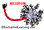
There are many type of neurons. They vary in size from 4 microns (.004 mm) to 100 microns (.1 mm) in diameter. Their length varies from a fraction of an inch to several feet.
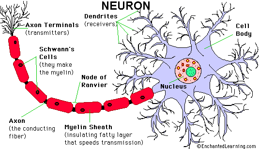
There are different types of neurons. They all carry electro-chemical nerve signals, but differ in structure (the number of processes, or axons, emanating from the cell body) and are found in different parts of the body.
- Sensory neurons or Bipolar neurons carry messages from the body's sense receptors (eyes, ears, etc.) to the CNS. These neurons have two processes. Sensory neuron account for 0.9% of all neurons. (Examples are retinal cells, olfactory epithelium cells.)
- Motoneurons or Multipolar neurons carry signals from the CNS to the muscles and glands. These neurons have many processes originating from the cell body. Motoneurons account for 9% of all neurons. (Examples are spinal motor neurons, pyramidal neurons, Purkinje cells.)
- Interneurons or Pseudopolare (Spelling) cells form all the neural wiring within the CNS. These have two axons (instead of an axon and a dendrite). One axon communicates with the spinal cord; one with either the skin or muscle. These neurons have two processes. (Examples are dorsal root ganglia cells.)
GLIAL CELLS
Glial cells make up 90 percent of the brain's cells. Glial cells are nerve cells that don't carry nerve impulses. The various glial (meaning "glue") cells perform many important functions, including: digestion of parts of dead neurons, manufacturing myelin for neurons, providing physical and nutritional support for neurons, and more. Types of glial cells include Schwann's Cells, Satellite Cells, Microglia, Oligodendroglia, and Astroglia. Neuroglia (meaning "nerve glue") are the another type of brain cell. These cells guide neurons during fetal development.
| The Spinal Cord |
 The spinal cord is a bundle of nerves that connects the brain to other parts of the body. It is protected by a series of doughnut-shaped bones called vertebrae, which surround the spinal cord. The human spinal cord is about 43-45 cm long and approximately as wide as a human finger. There are 13,500,000 neurons that transmit electro-chemical signals in the spinal cord. The cord weighs aproximately 35 grams. The vertebral column (bones) that supports it is about 70 cm long and has 31 segments and 31 pairs of spinal nerves. Spinal Cord Vertebrae
 - 7 cervical (neck) segments
- 12 thoracic segments
- 5 lumbar segments
- 5 sacral segments
- 4 fused coccygeal segment
| The Brain |
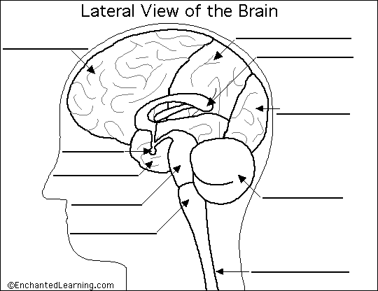
| Cerebellum - the part of the brain below the back of the cerebrum. It regulates balance, posture, movement, and muscle coordination. Corpus Callosum - a large bundle of nerve fibers that connect the left and right cerebral hemispheres. In the lateral section, it looks a bit like a "C" on its side. Frontal Lobe of the Cerebrum - the top, front regions of each of the cerebral hemispheres. They are used for reasoning, emotions, judgment, and voluntary movement. Medulla Oblongata - the lowest section of the brainstem (at the top end of the spinal cord); it controls automatic functions including heartbeat, breathing, etc. Occipital Lobe of the Cerebrum - the region at the back of each cerebral hemisphere that contains the centers of vision and reading ability (located at the back of the head). | Parietal Lobe of the Cerebrum - the middle lobe of each cerebral hemisphere between the frontal and occipital lobes; it contains important sensory centers (located at the upper rear of the head). Pituitary Gland - a gland attached to the base of the brain (located between the Pons and the Corpus Callosum) that secretes hormones. Pons - the part of the brainstem that joins the hemispheres of the cerebellum and connects the cerebrum with the cerebellum. It is located just above the Medulla Oblongata. Spinal Cord - a thick bundle of nerve fibers that runs from the base of the brain to the hip area, running through the spine (vertebrae). Temporal Lobe of the Cerebrum - the region at the lower side of each cerebral hemisphere; contains centers of hearing and memory (located at the sides of the head). |
No hay comentarios:
Publicar un comentario